Eye Investigations and Screening
Accurate treatment depends on accurate measurement. Our investigation unit provides the imaging and testing that establish exactly what is happening inside the eye.
WHAT WE MEASURE
The tests we run
None of these is painful and none involves an injection.
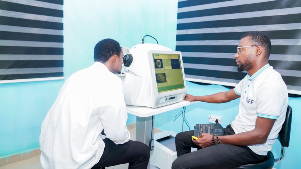
Photographs the retina, optic nerve and drainage angle in cross-section, layer by layer. You sit and look at a target. It is the single most useful scan in glaucoma and retinal disease.
Maps how much side vision you still have. You press a button each time a light appears, a few minutes per eye. This is the test that tracks whether glaucoma is stable or advancing.
Measures the thickness of the cornea, which changes how your pressure readings should be interpreted. A thin cornea can hide a dangerously high true pressure.
A small contact lens on a numbed eye lets the ophthalmologist see the drainage angle directly. It tells us which type of glaucoma you have, and that decides the treatment.
Measures the exact length and curvature of the eye so the right lens implant can be chosen before cataract surgery. Getting this measurement right is what decides your vision afterwards.
Sees behind a cataract too dense to look through. It finds retinal detachment, bleeding or a tumour in an eye whose view is completely blocked.
A photograph of the back of the eye, kept on record. It is how we show you what we can see, and how we compare this year against last.
Checks how well you distinguish colours. Loss of colour vision can be an early sign of optic nerve disease, and it matters for some occupations.
A simple grid you look at with one eye covered. Lines that appear bent, broken or missing point to a problem at the macula, the centre of the retina.
Why investigation matters
An eye examination tells us a great deal, but not everything. Investigation puts numbers and images to what the eye looks like: the thickness of the cornea, the shape of the drainage angle, the layers of the retina, and the exact length of the eye. Those measurements are what turn an impression into a diagnosis, and what let us tell later whether a condition is stable or getting worse.
They also become your baseline. A single visual field or OCT scan says far less than the comparison between this year’s and last year’s.
Which tests go with which condition
For glaucoma we normally run four together: OCT of the optic nerve, perimetry to map the visual field, pachymetry to interpret the pressure correctly, and gonioscopy to see the drainage angle. Each answers a different question, and glaucoma cannot be properly staged without all four.
Before cataract surgery we use biometry to measure the eye for its lens implant, and ocular ultrasound where the cataract is too dense to see through, so that we know the retina behind it is healthy before we operate.
For the retina we use colour fundus photography, posterior OCT, B scan ultrasound, and where the macula is involved, colour vision testing and the Amsler grid.
What the tests involve
None of these tests is painful, and none involves an injection.
OCT photographs the layers of the retina and optic nerve in cross-section; you simply sit and look at a target. Perimetry maps your field of vision; you press a button each time you see a light appear, and it takes a few minutes per eye. Pachymetry measures corneal thickness, which affects how your eye pressure readings should be interpreted. Gonioscopy uses a small contact lens on a numbed eye to look at the drainage angle. Biometry and ocular ultrasound measure the eye to plan cataract surgery, and to see behind a cataract too dense to look through.
Several of these tests need the pupil dilated, which blurs your vision for a few hours. Bring someone with you, and do not plan to drive yourself home.
Our screening programmes
Diabetic retinopathy screening. Everyone living with diabetes needs the retina examined every year, whether or not their sight has changed. We screen using colour fundus photography, OCT and, where the view is obscured, B scan ultrasound. Diabetic retinopathy is treatable when it is found early and very difficult to reverse once it is not.
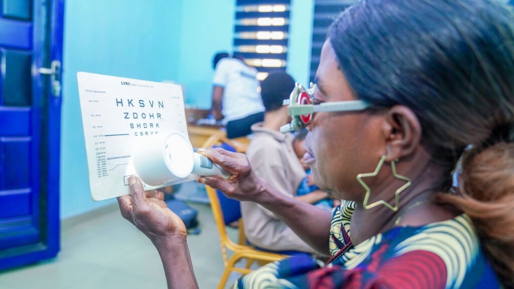
School eye health programme. We screen pupils and students at every level, from primary through junior and senior secondary to tertiary institutions. A child who cannot see the board is often labelled slow, inattentive or disruptive when the real problem is an uncorrected refractive error, and a pair of glasses settles it.
Occupational screening. We carry out fitness to drive and workplace vision screening for individuals, transport operators and companies.
Why screening matters
Glaucoma and diabetic eye disease both take sight away silently, long before a person notices anything wrong. By the time vision has changed, damage is usually well established and some of it is permanent. Screening is how both are caught while the sight can still be saved.
In children the same principle applies for a different reason. Uncorrected refractive error is one of the most common and most easily missed reasons for a child falling behind at school. That is why our school programme exists, and why we recommend an eye test before a child starts school.
Arranging a screening for your school, church or organisation
We run screening programmes for primary schools, junior and senior secondary schools and tertiary institutions, as well as occupational screening for drivers and company staff.
If you are responsible for a school, a workplace, a church or a community group and would like to arrange a screening, contact the hospital and ask for the investigation unit. Tell us roughly how many people are involved and where you are, and we will work out what is practical.
Book an appointment
Come and see our eye care team at St Joseph’s Eye Hospital, Mgbirichi. Emergency line (+234) 706 313 0156.